Pelvic Venous Insufficiency: Causes, Symptoms & Treatment
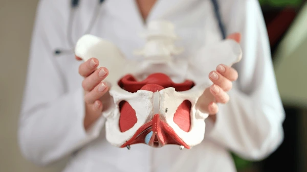
By Maria Shafiq | Founder & Health Research Writer, The Veins Vercel | Updated: 18 September 2026
Medical Disclaimer: The information provided in this article is for educational and informational purposes only and does not constitute medical advice. Always consult a qualified healthcare provider regarding any medical condition or treatment.
Introduction
Healthy blood circulation relies on an intricate network of blood vessels. Veins carry deoxygenated blood back to the heart against the pull of gravity. However, veins in the lower abdomen can struggle to maintain efficient upward flow. When this happens, understanding pelvic venous insufficiency becomes essential for protecting long-term vascular wellness.
In healthy vessels, tiny one-way valves prevent blood from flowing backward. If these valves weaken or fail, blood begins to pool in the pelvic basin. Consequently, surrounding veins stretch, engorge, and become varicose. Many individuals experience chronic, unexplained pelvic heaviness without knowing the vascular origin.
Historically, clinicians referred to this condition primarily as pelvic congestion syndrome. Today, vascular specialists recognize it as a broader spectrum of chronic venous disease. Living with pelvic venous insufficiency can disrupt daily movement, work, and personal comfort. Fortunately, evidence-based lifestyle adjustments and modern medical care can support venous health. This comprehensive guide explores the causes, symptoms, and practical management strategies for optimal pelvic vein function.
What Is Pelvic Venous Insufficiency?
Clinically, pelvic venous insufficiency occurs when retrograde blood flow develops in the ovarian or internal iliac veins [Source: National Institutes of Health]. In a balanced circulatory system, veins maintain low internal pressure. They rely on functional valves to direct blood upward toward the inferior vena cava and renal veins.
+-----------------------------------------------------------------------------------+
|                     NORMAL VS. INSUFFICIENT PELVIC VEIN DYNAMICS                  |
+-----------------------------------------------------------------------------------+
  HEALTHY VENOUS RETURN:
  - Competent Valves Close Tightly --> Retrograde Flow Blocked
  - Upward Hemodynamic Transit --> Normal Low Luminal Pressure
  - Flexible, Narrow Pelvic Vein Walls (< 5 mm diameter)
                                         vs.
  PELVIC VENOUS INSUFFICIENCY:
  - Incompetent or Absent Valves --> Gravity-Driven Reflux Occurs
  - Venous Blood Pools in Lower Pelvis --> Sustained Venous Hypertension
  - Chronic Engorgement --> Dilated, Tortuous Varicose Veins (> 5-6 mm)
+-----------------------------------------------------------------------------------+
However, when valves become incompetent, gravity pulls blood downward. As a result, blood pools in dependent pelvic venous reservoirs. This continuous backward pooling causes sustained venous hypertension within the pelvic floor. Over time, the affected pelvic vein walls stretch, widen, and become tortuous. When veins expand beyond 5 to 6 millimeters in diameter, they form varicose veins pelvis networks around pelvic organs.
Moreover, the condition involves both mechanical and hormonal factors. Estrogen naturally acts as a vascular smooth muscle relaxant. Therefore, elevated hormone levels can weaken vein walls and promote venous dilation. As blood flow stagnates, surrounding tissues experience reduced oxygen delivery. To understand how venous pooling contrasts with arterial wall resistance, review our clinical comparison of Arterial Stiffness vs Venous Insufficiency.
In addition, research indicates that pelvic venous hypertension triggers local neurovascular inflammation [Source: PubMed Central]. Stretched vein walls stimulate unmyelinated nerve fibers, transmitting dull, persistent pain signals to the spinal cord. Consequently, chronic discomfort often persists until venous pressure decreases.
Causes and Risk Factors
The development of pelvic venous insufficiency is multifactorial. Both anatomical variations and physiological stressors contribute to valve failure and venous dilation.
+-----------------------------------------------------------------------------------+
|                        PATHOPHYSIOLOGICAL ETIOLOGY TREE                           |
+-----------------------------------------------------------------------------------+
  Hormonal Influence (Estrogen-Mediated Vein Wall Relaxation)
                                    |
  Mechanical Compression (May-Thurner or Nutcracker Anatomical Variations)
                                    |
  Multiparity (Gestational Blood Volume Expansion & Pelvic Floor Strain)
                                    |
                                    v
  Venous Valve Incompetence & Reflux in Ovarian and Internal Iliac Veins
                                    |
                                    v
  Venous Stasis, Microvascular Congestion, and Pelvic Floor Heaviness
+-----------------------------------------------------------------------------------+
Primary Anatomical and Physiological Causes
-
Absence or incompetence of venous valves: Approximately 15% of women naturally lack valves in their left ovarian vein. Without functional valves, gravity forces blood to flow backward into the pelvis.
-
Mechanical venous compression: Conditions like May-Thurner syndrome compress the left common iliac vein between the spine and the right iliac artery. Similarly, Nutcracker phenomenon compresses the left renal vein between the aorta and mesenteric artery, forcing blood backward into pelvic tributaries.
-
Structural vein wall weakness: Genetic variations in extracellular collagen and elastin can reduce vein wall resilience. Over time, normal vascular pressures cause these susceptible vessels to stretch excessively.
Key Risk Factors
-
Multiple pregnancies (multiparity): During pregnancy, maternal blood volume increases by up to 50%. In addition, the growing uterus compresses pelvic outflow vessels, placing immense stress on venous valves.
-
Hormonal fluctuations: High circulating levels of estrogen and progesterone soften vascular smooth muscle. Therefore, premenopausal women experience higher rates of venous distension.
-
Prolonged standing or sitting: Occupations requiring long periods on one’s feet impede venous return. Gravity promotes continuous pooling in lower pelvic vascular beds.
-
Family history of venous disorders: Having relatives with chronic venous insufficiency or leg varicosities increases individual susceptibility [Source: Society for Vascular Surgery].
Common Symptoms
The clinical presentation of pelvic venous insufficiency varies among individuals. However, symptoms share a consistent vascular pattern related to posture and gravity.
+-----------------------------------------------------------------------------------+
|               COMMON CLINICAL MANIFESTATIONS & DIURNAL PATTERNS                   |
+-----------------------------------------------------------------------------------+
  Morning (Supine Rest):
  - Minimal to No Discomfort (Gravity Assists Overnight Venous Drainage)
                                         |
                                         v
  Midday to Evening (Upright Ambulation / Standing):
  - Progressive Venous Reflux and Stasis
  - Deep, Non-Cyclical Aching in the Lower Pelvis and Sacrum
  - Sensation of Heavy Fullness or Pressure in the Pelvic Floor
  - Visible Vulvar, Perineal, or Posterior Upper-Thigh Varicose Veins
+-----------------------------------------------------------------------------------+
Hallmark Symptoms of Pelvic Venous Congestion
-
Dull, aching pelvic pain: Patients typically report a persistent, non-cyclical ache deep in the lower abdomen or lower back. The discomfort is rarely sharp, presenting instead as a heavy pressure.
-
Postural worsening: Symptoms worsen significantly after standing or sitting upright for extended periods. Conversely, lying flat with elevated hips often provides noticeable relief.
-
Diurnal variation: Discomfort is usually minimal upon waking in the morning. However, pain progressively intensifies toward the end of the day as blood pools.
-
Post-coital ache: Increased blood flow during sexual arousal can exacerbate vascular engorgement. Consequently, many individuals experience a dull, throbbing ache that lasts for hours after intercourse.
-
Atypical varicose veins: Dilated veins may appear in unexpected locations, including the vulva, buttocks, and upper posterior thighs.
-
Bladder and bowel irritability: Engorged veins can press against adjacent pelvic organs. This pressure can cause a frequent urge to urinate or discomfort during bowel movements.
Furthermore, chronic vascular pooling can disrupt systemic microcirculation in connected areas. For insights into how poor upper-body circulation manifests, explore our guide on Poor Circulation in Head and Neck.
Diagnosis Methods
Accurate diagnosis of pelvic venous insufficiency requires careful vascular imaging. Because pelvic veins sit deep within the abdomen, routine physical examinations may appear entirely normal. Therefore, vascular specialists utilize targeted non-invasive and catheter-based imaging tools [Source: PubMed Study].
+-----------------------------------------------------------------------------------+
|                        DIAGNOSTIC WORKUP CASCADE                                  |
+-----------------------------------------------------------------------------------+
  Step 1: Pelvic & Transvaginal Duplex Ultrasound (Initial Non-Invasive Screen)
          - Measures ovarian vein diameter (> 5-6 mm)
          - Detects reversed or turbulent venous flow using Doppler imaging
                                    |
                                    v
  Step 2: Cross-Sectional Imaging (Contrast CT or Magnetic Resonance Venography)
          - Visualizes complex pelvic varicosities and venous anatomy
          - Screens for extrinsic vascular compression (Nutcracker / May-Thurner)
                                    |
                                    v
  Step 3: Transjugular / Transfemoral Catheter Venography (Gold Standard)
          - Confirms reflux under fluoroscopy
          - Measures exact intravenous pressure gradients before intervention
+-----------------------------------------------------------------------------------+
Primary Diagnostic Tools
-
Pelvic and transvaginal duplex ultrasound: Duplex ultrasound serves as the preferred initial screening modality. Specialists use color Doppler to assess flow direction in ovarian and internal iliac veins. A vein diameter exceeding 5 to 6 millimeters paired with reversed flow suggests venous incompetence.
-
Magnetic resonance venography (MRV): MRV provides high-resolution, three-dimensional views of pelvic vascular architecture without ionizing radiation. It clearly identifies engorged venous plexuses and evaluates compression points.
-
Computed tomography venography (CTV): Contrast-enhanced CT scans provide rapid, detailed mapping of pelvic veins. Furthermore, CTV effectively excludes other abdominal and pelvic conditions.
-
Catheter-directed venography: Clinicians consider catheter venography the gold standard diagnostic tool. During this minimally invasive procedure, a specialist inserts a thin catheter into the venous system under fluoroscopic guidance. Injecting contrast dye allows direct visualization of reflux, valve failure, and venous pooling in real time.
Treatment Options
Modern clinical management for pelvic venous insufficiency emphasizes restoring healthy pelvic hemodynamics. Physicians tailor therapies to symptom severity and individual anatomical findings.
+-----------------------------------------------------------------------------------+
|                      PELVIC VEIN TREATMENT SPECTRUM                               |
+-----------------------------------------------------------------------------------+
  CONSERVATIVE & LIFESTYLE CARE:
  - Pelvic floor physical therapy to enhance muscular pump function
  - External medical-grade compression shorts or garments
  - Position adjustments (frequent leg and hip elevation)
                                         |
  PHARMACOLOGICAL MANAGEMENT:
  - Phlebotonics (micronized purified flavonoid fractions) to support vein tone
  - Hormonal modulation (progestins or GnRH agonists) to suppress estrogenic dilation
                                         |
  MINIMALLY INVASIVE ENDOVASCULAR PROCEDURES:
  - Catheter-directed coil embolization with sclerosant agents
  - Venous stenting for relieving iliac or renal compression points
+-----------------------------------------------------------------------------------+
Conservative and Medical Management
Conservative approaches represent the first line of care for mild symptoms. Specialized pelvic floor physical therapy may help coordinate muscular support around deep veins. Strengthening the pelvic floor muscles encourages natural venous pumping during daily movement.
In addition, physicians may recommend medications to help manage vascular symptoms. Micronized purified flavonoid fractions (MPFF) are venactive compounds that research suggests may support venous tone and reduce capillary leakage. Hormonal therapies, such as medroxyprogesterone acetate or GnRH agonists, temporarily suppress ovarian estrogen production. By reducing circulating estrogen, these therapies may decrease vein wall distension and ease pelvic congestion.
Minimally Invasive Endovascular Interventions
When conservative methods provide insufficient relief, endovascular pelvic vein treatment offers a targeted solution. Interventional radiologists and vascular surgeons perform these procedures on an outpatient basis:
-
Pelvic vein embolization: Specialists access the venous system through a tiny puncture in the groin or neck. Guided by fluoroscopic imaging, the physician navigates a catheter into the refluxing ovarian or internal iliac veins. Small metallic coils and liquid sclerosants are placed to close off the incompetent, pooling vessels. Blood naturally reroutes into healthy, functional veins, relieving deep congestion.
-
Venous stenting: If May-Thurner syndrome or Nutcracker phenomenon is present, closing veins alone may not solve the problem. Instead, specialists place a self-expanding metal stent across the compressed vein. This restores an open outflow channel and lowers pelvic venous pressure.
7 Prevention Strategies for Pelvic Circulation
Proactive lifestyle habits play a meaningful role in supporting healthy pelvic circulation. While lifestyle changes cannot repair damaged vein valves, they may help minimize venous pooling and support vessel elasticity.
+-----------------------------------------------------------------------------------+
|               7 PILLARS OF DAILY PELVIC VASCULAR SUPPORT                          |
+-----------------------------------------------------------------------------------+
  1. Regular Aerobic Activity  --> Activates lower-limb and pelvic muscle pumps
  2. Targeted Nutrition        --> Bioflavonoids & nitrates support endothelial health
  3. Ergonomic Posture Breaks  --> Breaks up gravitational venous stagnation
  4. Daily Legs-Up Elevation   --> Mobilizes pooled venous blood toward the heart
  5. Consistent Hydration      --> Prevents blood viscosity elevation
  6. Pelvic Floor Exercise     --> Builds gentle muscular tone around deep veins
  7. Supportive Compression    --> Stabilizes superficial perineal vascular beds
+-----------------------------------------------------------------------------------+
1. Engage in Regular, Low-Impact Aerobic Exercise
Physical activity stimulates circulation throughout the entire body. When you walk, swim, or cycle, rhythmic skeletal muscle contractions compress nearby veins. This muscular pump pushes blood upward, reducing venous pooling in the lower pelvis. The American Heart Association recommends at least 150 minutes of moderate-intensity aerobic physical activity per week [Source: American Heart Association].
2. Nourish Your Vascular Lining with Nitrates and Bioflavonoids
What you eat influences vascular tone and vessel wall elasticity. Incorporating foods rich in bioflavonoids—such as citrus fruits, berries, and dark leafy greens—may support vein wall structural stability.
Furthermore, dietary nitrates found in beetroot and arugula promote endogenous nitric oxide production, which supports vascular compliance. To explore how nitrate-rich nutrition benefits blood vessel function, read our practical overview of Nitric Oxide Foods for Venous Compliance.
3. Take Frequent Ergonomic Movement Breaks
Prolonged, uninterrupted sitting or standing places continuous hydrostatic pressure on pelvic veins. Over several hours, gravity causes blood to settle in the pelvic floor.
To counteract this, set a timer to stand and walk for two minutes every hour. If your job requires standing, shift your weight frequently or use a small footstool to engage calf and pelvic muscles.
4. Practice Daily Hip and Leg Elevation
Elevating your lower extremities helps gravity work in your favor. At the end of the day, spend 10 to 15 minutes resting with your legs elevated on pillows or rested against a wall.
Placing a small cushion under your hips elevates the pelvis above heart level. This gentle tilt facilitates the drainage of pooled venous blood back into the central circulation.
5. Maintain Consistent Daily Hydration
Adequate hydration preserves healthy blood volume and prevents unnecessary blood thickening. When dehydration occurs, blood viscosity rises, creating greater resistance against vessel walls.
The World Health Organization emphasizes adequate fluid intake as a cornerstone of cardiovascular and metabolic vitality [Source: World Health Organization]. Drink clean water consistently throughout the day to keep circulation moving smoothly.
6. Practice Gentle Pelvic Floor Activation
The pelvic floor muscles form a supportive hammock beneath pelvic organs and blood vessels. Performing gentle, controlled pelvic floor contractions (Kegel exercises) can assist venous return.
However, avoid excessive straining or breath-holding during exercise. Straining elevates intra-abdominal pressure, which temporarily blocks venous outflow from the pelvis.
7. Utilize Supportive Compression Garments
Specialized maternity or pelvic compression shorts apply gentle external pressure across the lower abdomen, perineum, and upper thighs. This external support helps compress superficial dilated veins, preventing excessive distension during prolonged upright activity.
When to See a Doctor
While mild pelvic fullness can often be managed with daily lifestyle habits, persistent symptoms require medical attention. Chronic pelvic pain can stem from various conditions that warrant a formal medical evaluation.
+-----------------------------------------------------------------------------------+
|                        CLINICAL TRIAGE & RED FLAG SYMPTOMS                        |
+-----------------------------------------------------------------------------------+
  SCHEDULE A VASCULAR CONSULTATION:
  - Deep pelvic aching that progressively worsens throughout the day
  - Unexplained varicose veins on the vulva, buttocks, or upper inner thighs
  - Pelvic heaviness that consistently improves upon lying down flat
                                         vs.
  SEEK IMMEDIATE MEDICAL EVALUATION:
  - Sudden, severe, sharp abdominal or pelvic pain with fever
  - Visible blood in the urine or sudden unexplained rectal bleeding
  - Rapid, asymmetrical swelling and pain in one leg (potential deep vein thrombosis)
+-----------------------------------------------------------------------------------+
You should consult a qualified physician or vascular specialist if you experience:
-
Chronic pelvic heaviness lasting longer than six months without a clear gynecological cause.
-
Noticeable varicose veins appearing around the vulva, inner thigh, or buttocks.
-
Persistent pain after standing that interferes with your work or quality of life.
-
Painful intercourse followed by prolonged, throbbing pelvic discomfort.
A comprehensive evaluation ensures an accurate diagnosis and rules out other potential causes of pelvic pain, such as endometriosis or uterine fibroids.
Frequently Asked Questions (FAQ)
1. Is pelvic venous insufficiency the same as pelvic congestion syndrome?
Yes, the two terms describe the same underlying condition. Historically, clinicians used the term pelvic congestion syndrome to describe the cluster of symptoms caused by pooled blood. Today, medical organizations prefer pelvic venous insufficiency or pelvic venous disease. This updated name reflects the true vascular mechanism: incompetent vein valves and retrograde blood flow.
2. Can pelvic vein problems cause varicose veins in the legs?
Yes, refluxing pelvic veins can contribute directly to leg varicosities. When blood pools under high pressure in the pelvis, it seeks alternative escape routes. These escape points often channel blood into the upper thigh and saphenous vein tributaries. As a result, individuals may develop recurrent varicose veins on the back of the thighs or around the knee.
3. Does pregnancy permanently damage pelvic vein valves?
Pregnancy places substantial physical and hormonal demands on pelvic blood vessels. The combination of increased blood volume and high progesterone naturally stretches vein walls. While veins often contract after delivery, repeated pregnancies can permanently damage delicate valves in susceptible individuals. Over time, this cumulative stretching increases the risk of chronic venous reflux.
4. Can dietary changes alone cure pelvic venous insufficiency?
Dietary adjustments cannot physically repair incompetent or missing vein valves. However, a balanced diet rich in bioflavonoids and dietary nitrates may support healthy vascular tone and reduce systemic inflammation. Nutrition works best as an essential supportive component alongside regular movement, ergonomic habits, and medical guidance.
Conclusion
The vascular system of the pelvis is a delicate network responsible for draining deoxygenated blood from lower abdominal organs. When pelvic venous insufficiency occurs, damaged valves and sustained venous reflux allow blood to pool within the pelvic basin. This chronic stasis can lead to persistent heaviness, dull aching, and visible varicosities.
Fortunately, modern vascular medicine provides clear diagnostic pathways and effective management options. Non-invasive ultrasound and advanced venography help specialists pinpoint the precise location of venous reflux. For many individuals, daily preventative habits—such as regular walking, ergonomic breaks, and hip elevation—offer practical comfort. When necessary, minimally invasive catheter therapies can successfully close refluxing vessels and restore balanced hemodynamics.
Prioritizing your vascular health begins with recognizing the signs of poor pelvic circulation. By combining proactive lifestyle choices with professional medical care, you can support your veins and preserve lasting vitality.
Editorial Note
This article was independently researched and written based on current publicly available nutritional literature and does not represent the views of, and is not affiliated with, any external publication referenced.
References
-
National Institutes of Health. (2022). Pelvic Venous Disorders (PeVD): A Comprehensive Review. https://pmc.ncbi.nlm.nih.gov/articles/PMC9767768/
-
PubMed Central. (2022). The Clinical Problem of Pelvic Venous Disorders. https://pubmed.ncbi.nlm.nih.gov/35710285/
-
PubMed Study. (2022). Insights into Pelvic Venous Disorders and Clinical Diagnostics. https://pubmed.ncbi.nlm.nih.gov/36561941/
-
Society for Vascular Surgery. (2026). Pelvic Venous Disease Summit and Clinical Consensus. https://vascular.org/vascular-specialists/education-and-meetings/meetings/pelvic-venous-disease-summit-0
-
American Heart Association. (2024). Physical Activity Guidelines for Adults and Vascular Health. https://www.heart.org/en/healthy-living/fitness/fitness-basics/aha-recs-for-physical-activity-in-adults
-
World Health Organization. (2024). Physical Activity Factsheet and Global Health Recommendations. https://www.who.int/news-room/fact-sheets/detail/physical-activity