Non-Invasive Vascular Imaging: Plaque Detection Guide
By Maria Shafiq | Founder & Health Research Writer, The Veins Vercel | Oct 9, 2026
Medical Disclaimer: The information provided in this article is for educational and informational purposes only and does not constitute medical advice.Always consult a qualifiied healthcare provider regarding any medical condition or treatment.
Non-Invasive Vascular Imaging: A Guide to Plaque Detection
Non-invasive vascular imaging helps doctors examine blood vessels without passing a catheter through an artery. Depending on the test, it can show plaque buildup, narrowing, or changes in blood flow. However, some scans still require an intravenous injection of contrast dye.[radiologyinfo][radiologyinfo][pmc.ncbi.nlm.nih]
Newer technologies, including photon-counting CT and computer-assisted plaque analysis, may provide more detailed information. These advances are promising, but they do not mean that everyone needs a scan or that imaging can predict exactly when a heart attack will happen.[pmc.ncbi.nlm.nih][pubmed.ncbi.nlm.nih]
This guide explains the main tests, what recent research shows, and how doctors decide whether imaging is appropriate.
Key Takeaways
- Coronary CT angiography can detect calcified and non-calcified plaque in the arteries supplying the heart.[radiologyinfo][pmc.ncbi.nlm.nih]
- A coronary calcium scan measures calcified plaque. It does not directly measure non-calcified plaque or provide the same information as a CT angiogram.[pmc.ncbi.nlm.nih]
- Photon-counting CT can improve image detail and reduce some calcium-related distortion. Radiation benefits depend on the scanner and examination settings.[pmc.ncbi.nlm.nih]
- Ultrasound and MRI do not use ionising radiation, but they have different uses and limitations. They are not interchangeable with coronary CT.[radiologyinfo][pmc.ncbi.nlm.nih]
- Medical recommendation: Everyone’s symptoms, family history, kidney function, and risk factors are different. Ask a qualified healthcare professional whether imaging would change your care rather than choosing a scan based on an online article.[radiologyinfo][ahajournals]
- New chest pain or possible stroke symptoms need urgent medical assessment—not a routine screening appointment.[ahajournals][who]
What Is Non-Invasive Vascular Imaging?
Non-invasive vascular imaging is a group of tests used to examine arteries and veins without inserting an arterial catheter. These tests include ultrasound, CT-based scans, and magnetic resonance angiography, or MRA.[radiologyinfo][radiologyinfo][pmc.ncbi.nlm.nih]
This article focuses on arterial plaque, also called atherosclerosis.
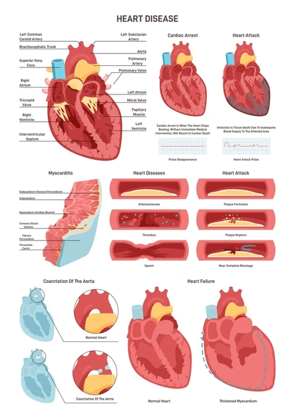
Plaque contains substances such as cholesterol, fat, and calcium. It builds up within artery walls and may eventually narrow the space through which blood flows.[radiologyinfo]
Different imaging tests answer different questions:
- Is plaque present?
- How much narrowing is there?
- Is blood flow affected?
- Are further tests or treatment needed?
No single scan provides every answer. Doctors choose tests according to the artery being examined, the symptoms, and the clinical question.[ahajournals][vascular]
Which Tests Can Detect Plaque?
Coronary Calcium Scan
A coronary artery calcium scan is a CT examination that measures calcified plaque in the heart’s arteries. It usually does not require contrast dye. Results are commonly reported as an Agatston score.[pmc.ncbi.nlm.nih]
The result may help clarify cardiovascular risk when a preventive treatment decision remains uncertain.
However, calcium scoring has limits. It does not directly measure non-calcified plaque, so a score of zero does not prove that every coronary artery is free of plaque.[pmc.ncbi.nlm.nih]
Coronary CT Angiography
Coronary CT angiography, often shortened to CCTA, uses CT scanning and an IV injection of iodine-containing contrast material.
It can show:
- Coronary artery anatomy.
- Calcified and non-calcified plaque.
- The location and extent of artery narrowing.[radiologyinfo]
CCTA is an established diagnostic option for selected people with suspected coronary artery disease. It may be useful when symptoms, initial test results, and overall risk suggest that looking directly at the arteries would help.[radiologyinfo][ahajournals]
It is not a routine screening test for every person without symptoms.[pmc.ncbi.nlm.nih]
Vascular Ultrasound
Vascular ultrasound uses sound waves to examine accessible blood vessels and assess blood flow. It does not use ionising radiation, and standard duplex ultrasound usually does not require injected contrast.[pmc.ncbi.nlm.nih]
A carotid ultrasound examines the neck arteries supplying the brain. It does not directly show plaque in the coronary arteries.
Importantly, the U.S. Preventive Services Task Force recommends against screening the general adult population for carotid artery narrowing when there are no relevant neurological symptoms or history. The potential harms of screening and subsequent procedures outweigh the expected benefits in that population.[uspreventiveservicestaskforce]
Magnetic Resonance Angiography
MRA uses magnetic fields and radio waves to examine blood vessels. It does not use ionising radiation. Some examinations require contrast, while others can be performed without it.[radiologyinfo]
MRA can be useful for selected vascular problems, but suitability depends on the vessel, the clinical question, and factors such as implanted devices or difficulty tolerating the scanner.[radiologyinfo][vascular]
What Is New About Photon-Counting CT?
Photon-counting CT is a newer form of CT technology. Its detectors count individual X-ray photons and measure their energy, rather than first converting X-rays into visible light.[pmc.ncbi.nlm.nih]
This can improve the detail available in cardiovascular images.
One potential advantage is reduced calcium blooming. Blooming is an image distortion that makes calcium appear larger than it really is, sometimes making the artery look more narrowed.[pmc.ncbi.nlm.nih]
A 2025 systematic review included 11 human studies involving 800 patients. Overall, photon-counting CT showed potential to improve image quality at similar radiation doses, or maintain image quality while reducing exposure in some applications.[pmc.ncbi.nlm.nih]
However, the studies used different protocols, and several were small. The authors called for larger, standardised studies to confirm benefits in routine practice.[pmc.ncbi.nlm.nih]
The practical message is that photon-counting CT is a promising improvement—not a guarantee of a lower dose or a better outcome for every patient.
Can Imaging Identify Higher-Risk Plaque?
Some plaque features seen on CT are associated with a greater risk of future cardiovascular events. One example is low-attenuation plaque, which appears less dense on CT and can reflect lipid-rich plaque.[pubmed.ncbi.nlm.nih]
What SCOT-HEART Found
A secondary analysis of the SCOT-HEART trial examined 1,769 patients with stable chest pain. Over a median follow-up of 4.7 years, 41 experienced a fatal or non-fatal heart attack.[pubmed.ncbi.nlm.nih]
Patients whose low-attenuation plaque burden exceeded 4% had approximately 4.65 times the relative hazard of a subsequent heart attack compared with those below that threshold.[pubmed.ncbi.nlm.nih]
This needs careful interpretation:
- The participants already had stable chest pain; they were not a general screening population.
- The result describes a relative association, not an individual’s absolute probability.
- It does not identify exactly which plaque will rupture or when.
- The plaque analysis was not itself a trial proving that treatment based on this threshold prevents heart attacks.[pubmed.ncbi.nlm.nih]
The finding supports looking beyond narrowing alone. It does not justify scanning everyone.
What About AI and Inflammation Analysis?
Computer-Assisted Plaque Measurements
Computer analysis can help assess plaque volume and imaging characteristics. However, measurements depend on image quality, reconstruction methods, and analysis settings. They should not be presented as exact tissue measurements or as a replacement for clinical interpretation.[pmc.ncbi.nlm.nih]
A detailed report is useful only when its findings can be interpreted in the context of the patient’s overall health and treatment options.
Perivascular Fat Attenuation
Researchers have also studied the fat surrounding coronary arteries.
The perivascular fat attenuation index, or FAI, is a CT-based marker intended to capture changes associated with coronary inflammation. It is an indirect imaging marker—not a direct measurement of inflammatory cells inside a plaque.[thelancet]
The CRISP-CT study analysed two groups of patients undergoing clinically indicated coronary CT angiography. Higher FAI values were associated with increased cardiac mortality after adjustment for other risk factors.[thelancet]
However, this was an analysis of observational outcome data. It did not establish that choosing treatment according to FAI improves patient outcomes. Predicting risk and proving the benefit of a treatment strategy are different questions.[thelancet]
How Do Non-Invasive Scans Compare With Catheter Angiography?
Neither approach is always better. They serve different purposes.
| Test | Main information | Important limitation or consideration |
| Coronary calcium scan | Amount of calcified coronary plaque | Does not directly measure non-calcified plaque; uses radiation |
| Coronary CT angiography | Coronary anatomy, plaque, and narrowing | Uses radiation and IV contrast; image quality can be affected by motion or extensive calcium |
| Vascular ultrasound | Blood flow and structure in accessible vessels | Does not directly assess coronary plaque; image quality varies |
| MRA | Blood-vessel anatomy in selected areas | Takes longer than CT; some examinations need contrast and implant checks |
| Catheter coronary angiography | Detailed assessment of coronary narrowing; can allow treatment in the same session | Requires arterial access and has procedural risks |
These differences guide test selection. A fixed radiation or resolution figure would be misleading because equipment, protocols, and patient characteristics vary.[radiologyinfo][radiologyinfo][pmc.ncbi.nlm.nih][pmc.ncbi.nlm.nih][vascular]
CCTA is a diagnostic test: a blockage cannot be treated during the scan. Catheter angiography can allow diagnosis and treatment, such as stenting, during the same procedure. It remains important when urgent intervention is needed.[radiologyinfo]
Who May Benefit From Vascular Imaging?
Imaging is most helpful when the result is likely to change diagnosis, treatment, or follow-up.
A clinician may consider coronary imaging for selected people with:
- Stable chest discomfort and an appropriate likelihood of coronary artery disease.
- Unclear or inconclusive previous test results.
- New or worsening symptoms despite a previous normal stress test.
- Other specific clinical reasons to assess coronary anatomy.[radiologyinfo][ahajournals]
For people without symptoms, preventive assessment usually starts with medical history and factors such as blood pressure, cholesterol, diabetes, smoking, and family history. Calcium scoring may help in selected situations, but imaging should complement—not replace—this assessment.[pmc.ncbi.nlm.nih][who]
There is no general recommendation that every healthy adult should request CCTA, carotid ultrasound, or advanced inflammation analysis.
Risks and Limitations
Radiation Exposure
CT-based tests use ionising radiation. The dose varies by examination, scanner, protocol, and patient characteristics. Ultrasound and MRI do not use ionising radiation.[radiologyinfo][radiologyinfo][pmc.ncbi.nlm.nih]
The question is whether the expected diagnostic benefit justifies the exposure—not whether a scan is completely risk-free.
Contrast and Kidney Function
CCTA usually requires iodinated contrast. People with impaired kidney function may need additional assessment or an alternative approach. Serious allergic reactions are uncommon but possible.[radiologyinfo]
Tell the imaging team about kidney disease, previous contrast reactions, pregnancy, and current medicines.
Image Quality
Irregular heart rhythms, movement, body size, and extensive calcification can affect CCTA quality. Some patients receive medicines to help obtain clearer images, depending on their circumstances.[radiologyinfo]
Incidental Findings and Further Testing
Scans may reveal findings unrelated to the original question, such as lung nodules. Some require follow-up, while others do not. This can add cost, uncertainty, or further testing.[pmc.ncbi.nlm.nih]
Access and Cost
Photon-counting scanners involve substantial equipment costs. The 2025 systematic review noted that clear evidence of cost-effectiveness was not yet available.[pmc.ncbi.nlm.nih]
The newest scanner is therefore not automatically the most appropriate choice.
What Happens If a Scan Finds Plaque?
Finding plaque does not automatically mean that you need a stent.
Your clinician will consider the extent of disease, symptoms, overall cardiovascular risk, and whether blood flow is affected. Treatment may include lifestyle support and medicines to manage cholesterol, blood pressure, or diabetes.[ahajournals][who]
Practical measures include:
- Avoiding tobacco.
- Staying physically active at an appropriate level.
- Following a balanced eating pattern.
- Taking prescribed medicines consistently.
- Managing blood pressure, diabetes, and cholesterol.[who]
A scan provides information. Reducing risk depends on what happens after the result.
When to Seek Urgent Medical Help
Do not wait for an outpatient scan if you develop possible heart attack or stroke symptoms.
Seek emergency care for:
- New chest pressure, tightness, or discomfort, especially with breathlessness, sweating, nausea, or faintness.
- Sudden facial drooping, one-sided weakness, or difficulty speaking.
- Sudden loss of vision, severe imbalance, or other possible stroke symptoms.[ahajournals][who]
Contact your local emergency service. Symptoms can vary, and you do not need every warning sign before getting help.
Frequently Asked Questions
Is non-invasive vascular imaging completely needle-free?
Not always. Standard ultrasound and coronary calcium scans generally do not require an IV injection. CCTA requires IV contrast, and some MRI examinations also use injected contrast.[radiologyinfo][radiologyinfo][pmc.ncbi.nlm.nih][pmc.ncbi.nlm.nih]
Can a CT scan detect plaque before an artery is severely narrowed?
Yes. CCTA can detect non-obstructive plaque, including non-calcified plaque. However, finding plaque does not reveal exactly when or whether a future heart attack will occur.[pubmed.ncbi.nlm.nih][pmc.ncbi.nlm.nih]
Does a calcium score of zero mean I have no plaque?
No. Calcium scoring detects calcified plaque, not all non-calcified plaque. Results must be interpreted alongside symptoms and other risk factors.[pmc.ncbi.nlm.nih]
Is photon-counting CT always lower in radiation?
No. It may improve dose efficiency, but the actual exposure depends on the protocol and the imaging task. Some high-resolution settings prioritise detail rather than the lowest possible dose.[pmc.ncbi.nlm.nih]
Should I have scans regularly to monitor plaque?
There is no single scanning schedule suitable for everyone. Follow-up imaging should answer a specific clinical question and be ordered when it is likely to influence care.[radiologyinfo][ahajournals]
Conclusion
Non-invasive vascular imaging can help doctors detect arterial plaque and investigate suspected vascular disease. CCTA is an established tool, while photon-counting CT and advanced image analysis may improve the detail available in selected cases.[radiologyinfo][pmc.ncbi.nlm.nih][ahajournals]
The safest approach is not to request the most advanced scan automatically. It is to choose the right test with a qualified clinician, understand its limitations, and use the result to guide appropriate care.
Editorial Note and Medical Disclaimer
This article provides general educational information. It does not diagnose vascular disease, recommend a particular scan for an individual, or replace professional medical advice.
Symptoms, medical history, kidney function, medicines, and cardiovascular risk differ between people. A qualified healthcare professional should decide whether imaging is appropriate and explain the benefits, limitations, and alternatives.
This draft draws on publicly available clinical guidance and research. The researchers and organisations cited do not endorse or have an affiliation with this website. No independent medical review is claimed.
Research and Clinical Sources
-
- Gulati M and colleagues. “2021 AHA/ACC/ASE/CHEST/SAEM/SCCT/SCMR Guideline for the Evaluation and Diagnosis of Chest Pain.” Circulation. 2021;144:e368–e454. DOI: 10.1161/CIR.0000000000001029.[ahajournals]
- RadiologyInfo.org. “Coronary CTA.” Reviewed June 15, 2026. Patient information from the American College of Radiology and Radiological Society of North America.[radiologyinfo]
- Williams MC and colleagues. “Low-Attenuation Noncalcified Plaque on Coronary Computed Tomography Angiography Predicts Myocardial Infarction: Results From the Multicenter SCOT-HEART Trial.” Circulation. 2020;141:1452–1462. DOI: 10.1161/CIRCULATIONAHA.119.044720.[pubmed.ncbi.nlm.nih]
- Rønning M, Johansen E, and Rusandu A. “Photon-counting CT versus energy-integrating detectors for cardiac imaging: a systematic review of evidence from in vivo human studies on image quality and radiation dose.” BMC Medical Imaging. 2025;25:295. DOI: 10.1186/s12880-025-01825-8.[pmc.ncbi.nlm.nih]
- Oikonomou EK and colleagues. “Non-invasive detection of coronary inflammation using computed tomography and prediction of residual cardiovascular risk: the CRISP CT study.” The Lancet. 2018;392:929–939. DOI: 10.1016/S0140-6736(18)31114-0.[thelancet]
- U.S. Preventive Services Task Force. “Asymptomatic Carotid Artery Stenosis: Screening.” February 2, 2021.[uspreventiveservicestaskforce]
- RadiologyInfo.org. “Magnetic Resonance Angiography.” Reviewed June 15, 2026.[radiologyinfo]
- World Health Organization. “Cardiovascular diseases (CVDs).” July 31, 2025.[who]
- Society for Vascular Surgery. “Computed Tomography Angiography (CTA) and Magnetic Resonance Angiography (MRA).”[vascular]
- “Use of cardiac CT and calcium scoring for detecting coronary plaque: implications on prognosis and patient management.” Published online in 2015.[pmc.ncbi.nlm.nih]