Arterial Stiffness vs. Venous Insufficiency: Key Differences
Medical Disclaimer: The information provided in this article is for educational and informational purposes only and does not constitute medical advice. Always consult a qualified healthcare provider regarding any medical condition or treatment.
1. Introduction: Understanding Arterial Stiffness vs. Venous Insufficiency in Systemic Hemodynamics
Understanding arterial stiffness vs venous insufficiency begins with the fundamental physics of the human circulatory system. The vascular tree operates as a dual-circuit closed hydraulic loop [Source: Journal of the American College of Cardiology].
Specifically, the high-pressure arterial system delivers oxygenated blood away from the left ventricle to peripheral capillary beds. In contrast, the low-pressure venous network channels deoxygenated blood back toward the right atrium against gravity.
Consequently, arterial stiffness vs venous insufficiency represents two entirely different failure modes within this circulatory network.
+-----------------------------------------------------------------------------------+
|                        SYSTEMIC CIRCULATORY HEMODYNAMICS                          |
+-----------------------------------------------------------------------------------+
  Left Ventricle (High Peak Systolic Pressure: ~120 mmHg)
       |
       v
  [Arterial Conduit System]  --> High-pressure distribution; requires elastic recoil
       |
       v
  Microvascular Capillary Bed --> Tissue nutrient exchange; pressure drops to ~20 mmHg
       |
       v
  [Venous Reservoir System]   --> Low-pressure capacitance; requires competent valves
       |
       v
  Right Atrium (Low Central Venous Pressure: ~0-5 mmHg)
+-----------------------------------------------------------------------------------+
High-Pressure Arterial Stiffness vs. Low-Pressure Venous Insufficiency
Arterial stiffness describes a structural loss of vascular elasticity in large conduit arteries, such as the aorta and carotid vessels. In healthy individuals, elastic arteries expand to cushion systolic pressure waves.
Subsequently, elastic recoil maintains steady diastolic blood flow to coronary and cerebral arteries. However, when arterial walls lose compliance, arteries act like rigid pipes.
In contrast, chronic venous insufficiency (CVI) stems from mechanical failure within the peripheral venous return system. Vein walls and delicate bicuspid valves fail to maintain forward, unidirectional flow.
As a result, gravity pulls blood backward into the lower extremities. This backward flow causes venous blood pooling and persistent ambulatory venous hypertension. To understand how microvascular lining integrity impacts fluid exchange in both circuits, explore our clinical analysis of endothelial glycocalyx integrity and vascular circulation.
Featured Snippet Direct Definition:
In arterial stiffness vs venous insufficiency, the primary difference involves vascular mechanics and circuit pressure. Arterial stiffness represents a loss of elastic compliance in high-pressure arteries, driving systolic hypertension. Venous insufficiency involves incompetent valves in low-pressure veins, causing blood pooling, swelling, and stasis in the lower extremities.
The Central Vascular Thesis
While both conditions impair systemic circulation, their cellular mechanisms diverge completely. Arterial stiffness reflects structural elastosis and medial collagen remodeling under high pressure.
In contrast, venous insufficiency stems from valvular reflux, venous dilation, and stagnant fluid leakage. Therefore, clinicians must distinguish between arterial inflow resistance and venous outflow pooling to design effective therapies.
Arterial Stiffness vs. Venous Insufficiency Diagnostic Comparison
| Clinical Marker | Arterial Stiffness (Conduit Arteriopathy) | Chronic Venous Insufficiency (Valvular Reflux) |
| System Operating Pressure | High-pressure circuit (Mean arterial pressure: 70–105 mmHg) | Low-pressure capacitance circuit (Resting venous pressure: 5–15 mmHg) |
| Primary Hemodynamic Defect | Accelerated pulse wave reflection; loss of aortic compliance | Incompetent bicuspid valves; sustained ambulatory venous hypertension |
| Primary Biomarker / Metric | Carotid-femoral Pulse Wave Velocity (cfPWV >10 m/s) | Retrograde venous reflux duration (>0.5 to 1.0 seconds on duplex) |
| Typical Pain Profile | Intermittent claudication; pain worsens with elevation | Heavy, dull aching; pain worsens with standing, improves with elevation |
| Physical Manifestations | Cold, pale extremities; diminished pulses; trophic skin changes | Brawny edema; varicose veins; stasis dermatitis; hemosiderin staining |
| Diagnostic Gold Standard | Non-invasive applanation tonometry (cfPWV assessment) | Venous Duplex Ultrasound with standardized distal augmentation |
2. Pathophysiology of Arterial Stiffness vs. Venous Insufficiency
To evaluate arterial stiffness vs venous insufficiency, physicians analyze the distinct cellular changes inside vessel walls [Source: Circulation Research]. High-pressure arterial remodeling differs fundamentally from low-pressure venous stagnation.
+-----------------------------------------------------------------------------------+
|                        VASCULAR PATHOPHYSIOLOGY PATHWAYS                          |
+-----------------------------------------------------------------------------------+
  ARTERIAL STIFFNESS CASCADE:
  Advanced Glycation End-Products (AGEs) --> Cross-link structural collagen
                     |
  Elastin Fragmentation & Calcinosis     --> Matrix metalloproteinases degrade elastin
                     |
  Accelerated Pulse Wave Velocity (PWV)  --> Wave reflections hit heart during systole
                     |
  Isolated Systolic Hypertension         --> Ventricular strain & reduced coronary flow
                                         vs.
  VENOUS INSUFFICIENT CASCADE:
  Valvular Incompetence & Reflux         --> Gravity drives retrograde blood pooling
                     |
  Ambulatory Venous Hypertension         --> Capillary bed experiences chronic backpressure
                     |
  Endothelial Gapping & Extravasation    --> Red blood cells and plasma leak into dermis
                     |
  Hemosiderin Deposition & Fibrosis      --> Inflammation sparks lipodermatosclerosis
+-----------------------------------------------------------------------------------+
Cellular Mechanisms of Arterial Stiffness: Loss of Elastic Compliance
The aorta dampens cardiac pressure spikes through the Windkessel effect. During systole, healthy elastic fibers inside the medial layer stretch to store roughly half of the ventricular stroke volume.
Subsequently, during diastole, these elastic walls recoil inward. This recoil propels blood forward, perfusing coronary arteries while the heart rests.
However, several biochemical processes damage this mechanism over time:
-
Collagen Cross-Linking: Circulating sugars bind to vascular structural proteins, producing Advanced Glycation End-products (AGEs). These compounds form rigid cross-links between collagen fibers within the arterial tunica media.
-
Elastin Fragmentation: Mechanical strain fatigues elastic lamellae over time. Furthermore, inflammatory matrix metalloproteinases (MMPs) degrade fragmented elastin scaffolds.
-
Medial Calcinosis: Calcium phosphate minerals deposit directly onto stiffened collagen matrix fibers. Consequently, the arterial wall loses its capacity to stretch.
-
Accelerated Wave Reflection: In supple arteries, forward systolic pulse waves travel slowly (~4–6 m/s). Consequently, the reflected wave returns to the central aorta during diastole, supporting coronary blood flow. In stiffened arteries, the wave accelerates (>10 m/s). As a result, the reflected wave arrives prematurely during late systole. This mistimed wave increases left ventricular afterload and spikes central systolic pressure.
Cellular Mechanisms of Venous Insufficiency: Valvular Reflux and Stasis
Unlike arteries, veins rely on external skeletal muscle contractions to return blood toward the heart. Because humans stand upright, gravity exerts heavy hydrostatic pressure on the lower extremities.
To overcome this pressure, the body utilizes the calf muscle pump. As the gastrocnemius contracts, it compresses deep veins, driving blood upward through one-way bicuspid valves.
Venous insufficiency develops when these delicate valves fail:
-
Valvular Incompetence: Chronic mechanical stress, prior blood clots, or genetic vein wall weakness dilates venous lumens. Consequently, delicate valve leaflets separate and fail to close.
-
Retrograde Reflux: When the calf muscle relaxes, blood falls backward through incompetent valves.
-
Ambulatory Venous Hypertension (AVH): During walking, a healthy calf pump lowers foot venous pressure from 90 mmHg to below 25 mmHg. In patients with incompetent valves, pressure remains above 60 to 80 mmHg throughout ambulation.
-
Microvascular Extravasation: High backpressure stretches microvascular capillaries in the lower legs. Consequently, endothelial junctions separate, allowing plasma proteins, fluid, and red blood cells to leak into the dermis. Macrophages break down leaked erythrocytes, releasing hemosiderin iron pigments that stain the skin dark brown. To review how cellular fluid balance preserves tissue structure, consult our guide on tissue hydration and cellular fluid balance.
Endothelial Shear Stress in Arterial Stiffness vs. Venous Insufficiency
Endothelial cells adapt directly to local hemodynamic shear stress:
-
Arterial Endothelial Changes: Pulsatile laminar shear stress stimulates endothelial nitric oxide synthase (eNOS). Nitric oxide (NO) diffuses into vascular smooth muscle, promoting vasodilation. However, arterial stiffening reduces wall compliance and generates disturbed flow. Consequently, eNOS expression falls, increasing vascular tone and platelet aggregation.
-
Venous Endothelial Changes: In venous insufficiency, stagnant, low-velocity flow activates endothelial adhesion molecules (ICAM-1, VCAM-1). These proteins recruit circulating white blood cells, which attach to valve leaflets and release inflammatory proteases that accelerate valve destruction.
3. Physical Symptoms of Arterial Stiffness vs. Venous Insufficiency
A structured physical examination easily distinguishes arterial stiffness vs venous insufficiency [Source: New England Journal of Medicine]. Clinicians inspect limb temperature, skin coloration, peripheral pulses, and edema patterns.
+-----------------------------------------------------------------------------------+
|                        PHYSICAL PRESENTATION CHECKLIST                            |
+-----------------------------------------------------------------------------------+
  Arterial Compromise (Inflow Failure):
  - Temperature: Cold to the touch; cutaneous hypothermia
  - Color: Dependent rubor; pallor upon limb elevation
  - Pulses: Weak, diminished, or completely absent pedal pulses
  - Edema: Minimal to absent; legs appear slender or atrophic
  - Pain Relief: Pain worsens with elevation; improves when legs dangle downward
                                         vs.
  Venous Stasis (Outflow Failure):
  - Temperature: Warm or normal skin temperature
  - Color: Brownish hemosiderin hyperpigmentation; cyanotic venous engorgement
  - Pulses: Readily palpable (frequently masked by overlying fluid edema)
  - Edema: Prominent, brawny, pitting or non-pitting edema of the ankle and calf
  - Pain Relief: Pain worsens with standing; improves when limbs elevate above heart
+-----------------------------------------------------------------------------------+
Lower Extremity Pain Patterns in Arterial Stiffness vs. Venous Insufficiency
The character and timing of leg discomfort separate arterial disease from venous disease:
-
Arterial Pain Characteristics: Patients experience intermittent claudication. During walking, contracting calf muscles require more oxygenated blood. Because rigid, narrowed arteries cannot dilate, muscle tissue suffers transient ischemia. Consequently, patients experience sharp, cramping pain that forces them to stop. Resting for several minutes restores blood flow, relieving discomfort. In advanced stages, patients experience ischemic rest pain in the toes at night. To find relief, patients hang their feet over the bed edge, using gravity to pull blood downward into ischemic tissues.
-
Venous Pain Characteristics: In contrast, venous insufficiency creates a dull ache, throbbing, and heavy fatigue. Pain worsens after prolonged standing or sitting because stagnant blood engorges the lower leg. Walking actually improves venous symptoms by activating the calf muscle pump. Furthermore, elevating the legs above heart level drains pooled blood, providing rapid pain relief.
Cutaneous Changes in Arterial Stiffness vs. Venous Insufficiency
Chronic vascular disease produces distinct skin changes:
+-----------------------------------------------------------------------------------+
|                        CUTANEOUS ULCER DIFFERENTIATION                            |
+-----------------------------------------------------------------------------------+
  Arterial Ischemic Ulcers:
  - Location: Distal pressure points; tips of toes, lateral malleolus, heel margins
  - Margins: Sharply "punched-out" edges; round, clean borders
  - Wound Bed: Pale, dry, gray, or necrotic base; minimal wound exudate
  - Surrounding Skin: Shiny, hairless, atrophic skin; cool with thick brittle nails
                                         vs.
  Venous Stasis Ulcers:
  - Location: Medial lower calf; directly over or above the medial malleolus
  - Margins: Irregular, sloping, shallow borders
  - Wound Bed: Moist, beefy red granulation tissue; heavy serosanguinous exudate
  - Surrounding Skin: Brown hemosiderin deposits; warm, brawny, fibrotic induration
+-----------------------------------------------------------------------------------+
Arterial disease starves dermal hair follicles and fibroblasts. Consequently, the skin becomes thin, shiny, and hairless, while toenails become thick and brittle. If an arterial ulcer develops, it forms a dry, pale, “punched-out” circular sore over distal pressure points like the toe tips or outer lateral ankle.
In contrast, chronic venous insufficiency causes stasis dermatitis. Extravasated red blood cells break down, staining the skin of the inner calf and ankle a characteristic rust-brown color.
Over time, chronic inflammation triggers lipodermatosclerosis, an induration where subcutaneous fat fibroses into dense scar tissue. This scarring narrows the lower ankle while the upper calf remains swollen, creating an inverted champagne bottle appearance. When venous ulcers develop, they appear around the medial malleolus as shallow, moist, weeping sores with irregular edges. To understand how epithelial barriers heal under inflammatory stress, read our analysis on epithelial and mucosal barrier integrity.
4. Distinct Causes of Arterial Stiffness vs. Venous Insufficiency
When evaluating the causes of arterial stiffness vs venous insufficiency, physicians identify distinct clinical risk profiles [Source: Hypertension]. Arterial stiffness reflects systemic vascular aging, whereas venous insufficiency stems from mechanical valvular trauma and gravitational strain.
+-----------------------------------------------------------------------------------+
|                        SYSTEMIC ETIOLOGICAL PROFILES                              |
+-----------------------------------------------------------------------------------+
  ARTERIAL STIFFNESS TRIGGERS:
  - Chronological Vascular Aging (Elastin fragmentation over decades)
  - Essential Hypertension (High shear stress accelerates wall remodeling)
  - Diabetes Mellitus (Hyperglycemia accelerates advanced glycation cross-linking)
  - Chronic Kidney Disease (Hyperphosphatemia drives medial calcification)
  - Dyslipidemia & Smoking (Endothelial injury accelerates vascular fibrosis)
                                         vs.
  VENOUS INSUFFICIENT TRIGGERS:
  - Familial Venous Incompetence (Inherited matrix metalloproteinase alterations)
  - Occupational Orthostasis (Sustained standing or seated immobility)
  - Prior Deep Vein Thrombosis (Post-thrombotic valvular destruction)
  - Pregnancy (Elevated progesterone relaxes veins; gravid uterus compresses cava)
  - Central Obesity (Increased intra-abdominal pressure opposes femoral return)
+-----------------------------------------------------------------------------------+
Risk Factors for Arterial Stiffness
Arterial stiffening develops through several well-documented metabolic and hemodynamic drivers:
-
Vascular Aging: With each heartbeat, the aorta expands and recoils over 100,000 times each day. Over decades, this mechanical fatigue fractures elastin polymers, shifting the pressure load onto rigid collagen.
-
Essential Hypertension: Chronic high blood pressure exerts high mechanical strain on vessel walls. In response, vascular smooth muscle cells synthesize dense collagen, accelerating wall stiffness.
-
Diabetes and Glucotoxicity: Elevated blood glucose accelerates the formation of advanced glycation end-products. These sugar complexes bind directly to vascular structural proteins, stiffening the arterial matrix.
-
Chronic Kidney Disease (CKD): Renal impairment disrupts calcium and phosphate homeostasis. Consequently, vascular smooth muscle cells deposit calcium hydroxyapatite directly into the arterial media (Mönckeberg sclerosis).
Risk Factors for Venous Insufficiency
In contrast, venous insufficiency develops from factors that compromise valvular mechanics:
-
Occupational Orthostasis: Occupations that demand prolonged standing (such as retail workers, nurses, and surgeons) maintain high hydrostatic pressure in lower extremity veins. Without regular calf muscle contractions, stagnant blood pools in the legs, stretching vein walls apart.
-
Prior Deep Vein Thrombosis (DVT): A blood clot inside deep veins triggers intense inflammation that scars delicate valve leaflets. Over time, up to 50% of DVT patients develop post-thrombotic syndrome, characterized by permanent valvular reflux and chronic swelling.
-
Pregnancy: Elevated progesterone relaxes vascular smooth muscle, increasing venous compliance. Furthermore, the expanding uterus compresses pelvic veins, impeding venous drainage from both legs.
-
Elevated Intra-Abdominal Pressure: Visceral adiposity compresses pelvic veins, elevating baseline venous pressure in the lower extremities.
5. Clinical Diagnosis of Arterial Stiffness vs. Venous Insufficiency
To differentiate arterial stiffness vs venous insufficiency, vascular specialists utilize non-invasive physiological testing and ultrasound imaging [Source: Journal of Vascular Surgery].
                  +-------------------------------------------------------------+
                  |               VASCULAR DIAGNOSTIC WORKUP                    |
                  +-------------------------------------------------------------+
                                                 |
                       +-------------------------+-------------------------+
                       |                                                   |
                       v                                                   v
        +-----------------------------+                     +-----------------------------+
        | Arterial Compliance Workup  |                     | Venous Hemodynamic Workup   |
        +-----------------------------+                     +-----------------------------+
        | - Carotid-Femoral PWV       |                     | - Duplex Ultrasound (Reflux)|
        | - Augmentation Index (AIx)  |                     | - Air Plethysmography (APG) |
        | - Ankle-Brachial Index (ABI)|                     | - Ambulatory Venous Pressure|
        | - Arterial Duplex Doppler   |                     | - Venous Filling Index (VFI)|
        +-----------------------------+                     +-----------------------------+
Testing Modalities for Arterial Stiffness and Venous Insufficiency
Vascular specialists deploy specific diagnostic tools based on the vascular bed:
-
Carotid-Femoral Pulse Wave Velocity (cfPWV): Considered the gold standard for measuring arterial stiffness. Technicians place pressure tonometers over the right common carotid and right femoral arteries. Electrocardiogram gating measures the transit time of the pulse wave across the measured distance. A velocity exceeding 10 meters per second confirms significant arterial stiffness and elevated cardiovascular risk.
-
Augmentation Index (AIx): Derived from pulse wave analysis, this test measures the percentage by which the reflected wave boosts central aortic systolic pressure. Higher values confirm premature wave reflections from stiff peripheral vascular beds.
-
Ankle-Brachial Index (ABI): A Doppler probe measures systolic blood pressure at the arm and the ankle. An ABI ratio below 0.90 confirms peripheral artery disease, whereas an ABI above 1.40 indicates rigid, non-compressible, calcified tibial arteries.
-
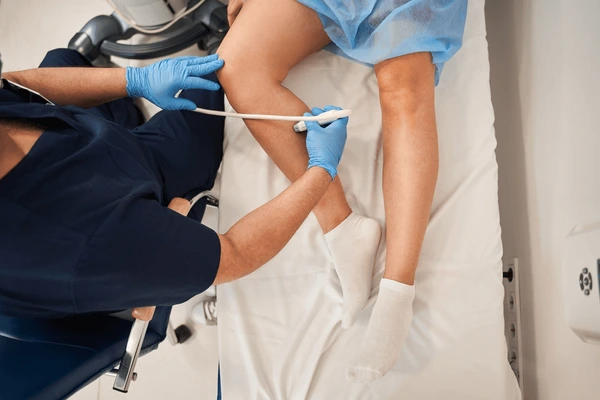
Venous Duplex Ultrasound with Reflux Protocol: The gold-standard imaging tool for diagnosing venous insufficiency. The patient stands upright to ensure maximum gravitational filling. Next, the sonographer compresses the calf and releases it, measuring backward flow:
-
In the superficial saphenous system, retrograde flow lasting longer than 500 milliseconds (0.5 seconds) confirms pathological reflux.
-
In deep femoral or popliteal veins, retrograde flow exceeding 1,000 milliseconds (1.0 second) confirms deep venous insufficiency.
-
-
Air Plethysmography (APG): Inflatable cuffs around the calf measure limb volume changes during postural shifts, quantifying the Venous Filling Index (VFI) and assessing calf muscle pump efficiency. To explore comprehensive biomarker panels that support cardiovascular assessment, review our guide on nutrient diagnostics and functional biomarker testing.
Diagnostic Testing Breakdown Matrix
| Diagnostic Test | Target Parameter | Clinical Purpose | Diagnostic Criteria / Findings |
| Carotid-Femoral PWV | Transit time of aortic pulse wave | Quantifies aortic stiffness directly | cfPWV >10 m/s confirms significant arterial stiffness |
| Ankle-Brachial Index (ABI) | Ratio of ankle to arm systolic pressure | Detects macrovascular flow obstruction | ABI <0.90 confirms PAD; ABI >1.40 confirms calcification |
| Augmentation Index (AIx) | Central aortic pressure wave reflection | Assesses peripheral wave reflection timing | Elevated AIx (>25–30%) confirms early reflected wave arrival |
| Venous Duplex Ultrasound | Duration of retrograde Doppler flow | Pinpoints specific incompetent valves | Retrograde reflux >0.5s (superficial) or >1.0s (deep) |
| Air Plethysmography (APG) | Dynamic calf volume variations (mL/s) | Measures ambulatory venous filling rate | High filling rate (>4 mL/s) confirms severe venous reflux |
6. Evidence-Based Treatments for Arterial Stiffness vs. Venous Insufficiency
Managing arterial stiffness vs venous insufficiency requires tailored clinical interventions. Treating arterial stiffness focuses on lowering central pressure and restoring endothelial compliance.
In contrast, treating venous insufficiency focuses on relieving gravitational backpressure and eliminating incompetent vein segments [Source: European Heart Journal].
                           +---------------------------------------+
                           |    SYSTEMIC VASCULAR MANAGEMENT       |
                           +---------------------------------------+
                                              |
        +-------------------------------------+-------------------------------------+
        |                                     |                                     |
        v                                     v                                     v
+-------------------------------+   +-------------------------------+   +-------------------------------+
|      Arterial Therapeutics    |   |     Venous Therapeutics       |   |      Interventional Care      |
+-------------------------------+   +-------------------------------+   +-------------------------------+
| - ACE Inhibitors & ARBs       |   | - Graduated Compression Hose  |   | - Endovenous Thermal Ablation |
| - Dihydropyridine CCBs        |   | - Scheduled Leg Elevation     |   | - Ultrasound Foam Sclerotherapy|
| - Dietary Inorganic Nitrates  |   | - Micronized Flavonoids (MPFF)|   | - Varithena Microfoam         |
| - High-Shear Aerobic Training |   | - Calf Muscle Conditioning    |   | - Surgical Bypass (Arterial)  |
+-------------------------------+   +-------------------------------+   +-------------------------------+
Therapeutic Protocols for Arterial Stiffness vs. Venous Insufficiency
Clinicians deploy distinct treatments based on vascular physiology:
-
Arterial Stiffness Therapies:
-
RAAS Inhibitors: ACE inhibitors and ARBs lower peripheral resistance. Furthermore, by blocking angiotensin II signaling, these drugs reduce vascular fibrosis and slow arterial wall remodeling.
-
Dihydropyridine Calcium Channel Blockers (CCBs): Medications like amlodipine relax arterial smooth muscle, delaying wave reflections and lowering central systolic pressure.
-
Dietary Nitrates: Concentrated beetroot extract and leafy greens provide inorganic nitrates that convert into nitric oxide. This supplemental nitric oxide dilates stiffened conduit arteries and improves flow-mediated dilation.
-
Aerobic Exercise Training: Regular aerobic exercise (brisk walking, cycling, or swimming) stimulates pulsatile blood flow across endothelial surfaces. This healthy shear stress activates eNOS, reducing arterial stiffness by up to 15% over three to six months.
-
-
Venous Insufficiency Therapies:
-
Graduated Compression Stockings: Compression socks deliver graded external pressure that is highest at the ankle (typically 20–30 mmHg) and decreases toward the knee. This pressure compresses superficial veins, restoring valve contact and speeding deep venous return. Caution: Clinicians must verify an ABI >0.8 prior to prescribing compression to ensure adequate arterial perfusion.
-
Scheduled Leg Elevation: Elevating the legs above heart level for 20 to 30 minutes three times daily drains pooled blood, lowering ambulatory venous hypertension.
-
Micronized Purified Flavonoid Fractions (MPFF): Formulations containing purified diosmin and hesperidin improve venous wall tone, reduce capillary leakage, and support lymphatic drainage.
-
Endovenous Thermal Ablation (EVLA / RFA): A vascular specialist inserts a thin catheter into an incompetent saphenous vein. Radiofrequency energy or laser heat seals the diseased vein shut. Consequently, blood reroutes into healthy deep veins with working valves.
-
Ultrasound-Guided Foam Sclerotherapy: Clinicians inject an irritant foam into tortuous varicose veins, closing the vessel and allowing the body to absorb it naturally.
-
Comparative Vascular Therapy Matrix
| Therapeutic Category | Arterial Stiffness Protocols | Venous Insufficiency Protocols | Clinical Rationale |
| First-Line Lifestyle | Aerobic exercise, low sodium, smoking cessation | Leg elevation, calf pump exercise, weight loss | Restores compliance (arterial); reduces backpressure (venous) |
| Mechanical Devices | Continuous Positive Airway Pressure (CPAP) | Graduated compression stockings (20–40 mmHg) | CPAP lowers nocturnal stress; stockings restore venous flow |
| First-Line Pharmacology | RAAS inhibitors, Dihydropyridine CCBs | Micronized Purified Flavonoid Fraction (MPFF) | Reduces collagen stiffness; improves venous tone |
| Interventional Therapy | Percutaneous balloon angioplasty and stenting | Endovenous thermal ablation (Laser / RF) | Re-establishes arterial inflow; seals leaking vein valves |
7. Red Flag Emergencies in Arterial Stiffness and Venous Insufficiency
While arterial stiffness vs venous insufficiency usually progress as chronic conditions, acute circulatory disruptions represent life-threatening emergencies.
+-----------------------------------------------------------------------------------+
|                         VASCULAR EMERGENCY RED FLAGS                              |
+-----------------------------------------------------------------------------------+
  ACUTE LIMB ISCHEMIA (Arterial Emergency):
  [The Six "P"s] --> Sudden onset of severe pain, pallor, pulselessness,
                     paresthesias (numbness), paralysis (weakness), and poikilothermia.
              |
  Requires immediate revascularization within 6 hours to prevent amputation.
                                         vs.
  ACUTE DEEP VEIN THROMBOSIS (Venous Emergency):
  [DVT Triad]    --> Rapid unilateral calf or thigh swelling, warmth, redness, and
                     tenderness, with potential sudden shortness of breath (PE).
              |
  Requires immediate diagnostic ultrasound and systemic anticoagulant therapy.
+-----------------------------------------------------------------------------------+
Acute Arterial Occlusion: The Six “P”s
When an embolus or thrombus suddenly occludes an artery, distal tissue suffers acute, severe ischemia. Patients present with the classic Six Ps:
-
Pain: Sudden, severe pain throughout the distal foot or leg.
-
Pallor: The limb turns chalky white or mottled.
-
Pulselessness: Complete loss of pedal pulses.
-
Poikilothermia: The foot feels ice-cold to the touch.
-
Paresthesias: Numbness or tingling sensations.
-
Paralysis: Inability to move the toes or foot.
Clinical Imperative: Patients displaying these signs must proceed to an emergency department immediately. Irreversible skeletal muscle necrosis occurs within six hours of complete arterial occlusion.
Acute Deep Vein Thrombosis (DVT)
In contrast, an acute venous clot obstructs blood leaving the limb. Patients develop sudden, unilateral swelling in the calf or thigh.
The leg feels warm, heavy, and tender to the touch. If a piece of the clot breaks free, it travels through the vena cava into the lungs, producing a life-threatening Pulmonary Embolism (PE).
Patients who experience sudden shortness of breath, pleuritic chest pain, or coughing up blood require emergency medical care immediately.
Selecting the Right Specialist
-
Consult a Cardiologist or Vascular Specialist: If you have elevated systolic blood pressure, abnormal pulse wave velocity, or exertional calf cramping (claudication).
-
Consult a Vascular Surgeon or Phlebologist: If you have prominent varicose veins, heavy swollen legs, skin pigmentation changes around the ankles, or non-healing leg wounds.
8. Frequently Asked Questions
Q1. Can you have both arterial stiffness and venous insufficiency at the same time?
Q2. Does elevating your legs help arterial stiffness?
Q3. How do doctors test pulse wave velocity?
Q4. Is high systolic blood pressure a sign of arterial stiffness?
Q5. Why do compression socks help venous insufficiency but hurt arterial disease?
9. Conclusion: Protecting Your Vascular Health
Editorial Note
This article was independently researched and written based on current publicly available peer-reviewed nutritional literature and does not represent the views of, and is not affiliated with, any external publication referenced.
References
-
Townsend, R. R., et al. (2015). Recommendations for Improving and Standardizing Vascular Research on Arterial Stiffness: A Scientific Statement From the American Heart Association. Hypertension, 66(3), 698–716. https://doi.org/10.1161/HYP.0000000000000033
-
Eberhardt, R. T., & Raffetto, J. D. (2014). Chronic venous insufficiency. Circulation, 130(4), 333–346. https://doi.org/10.1161/CIRCULATIONAHA.113.006898
-
Laurent, S., et al. (2006). Expert consensus document on arterial stiffness: methodological issues and clinical applications. European Heart Journal, 27(21), 2588–2605. https://doi.org/10.1093/eurheartj/ehl254
-
Bergan, J. J., et al. (2006). Chronic venous disease. New England Journal of Medicine, 355(5), 488–498. https://doi.org/10.1056/NEJMra055289
-
Vlachopoulos, C., et al. (2010). Prediction of cardiovascular events and all-cause mortality with arterial stiffness: a systematic review and meta-analysis. Journal of the American College of Cardiology, 55(13), 1318–1327. https://doi.org/10.1016/j.jacc.2009.10.061
-
Raffetto, J. D., & Khalil, R. A. (2008). Mechanisms of varicose vein formation and the role of matrix metalloproteinases in vein wall remodeling. Journal of Vascular Surgery, 47(3), 669–679. https://doi.org/10.1016/j.jvs.2007.09.033
-
Lurbe, E., et al. (2023). 2023 ESH Guidelines for the management of arterial hypertension. Journal of Hypertension, 41(12), 1874–2071. https://doi.org/10.1097/HJH.0000000000003480
-
Nicolaides, A. N., et al. (2018). Management of chronic venous disorders of the lower limbs: Guidelines according to scientific evidence. Part I. International Angiology, 37(3), 181–254. https://doi.org/10.23736/S0392-9590.18.03999-8
Comments (2)
Leg Numbness: Is It MS? 7 Causes Explained - The Veins Vercel
[…] Artery Disease involves atherosclerotic plaque buildup within lower extremity arteries. As the arterial lumen narrows, downstream tissue perfusion falls significantly. Ischemic leg […]
Tissue Hydration: Can Hyaluronic Acid Alter Aging - The Veins Vercel
[…] reach deep joint cartilage. Similarly, topical gels cannot restore the vascular lining of major arteries. Therefore, longevity researchers prioritize systemic hydration protocols over temporary surface […]