AI Brain Aneurysm Detection: How AI Supports Radiologists
Medical Disclaimer: The information provided in this article is for educational and informational purposes only and does not constitute medical advice. Always consult a qualified healthcare provider regarding any medical condition or treatment.
AI in Radiology: How AI Helps Doctors Detect Brain Aneurysms
A brain aneurysm is a bulge in a weakened blood vessel in the brain. Many aneurysms cause no symptoms and are found only during imaging for another reason. When one ruptures, it can cause bleeding around the brain and needs emergency care.
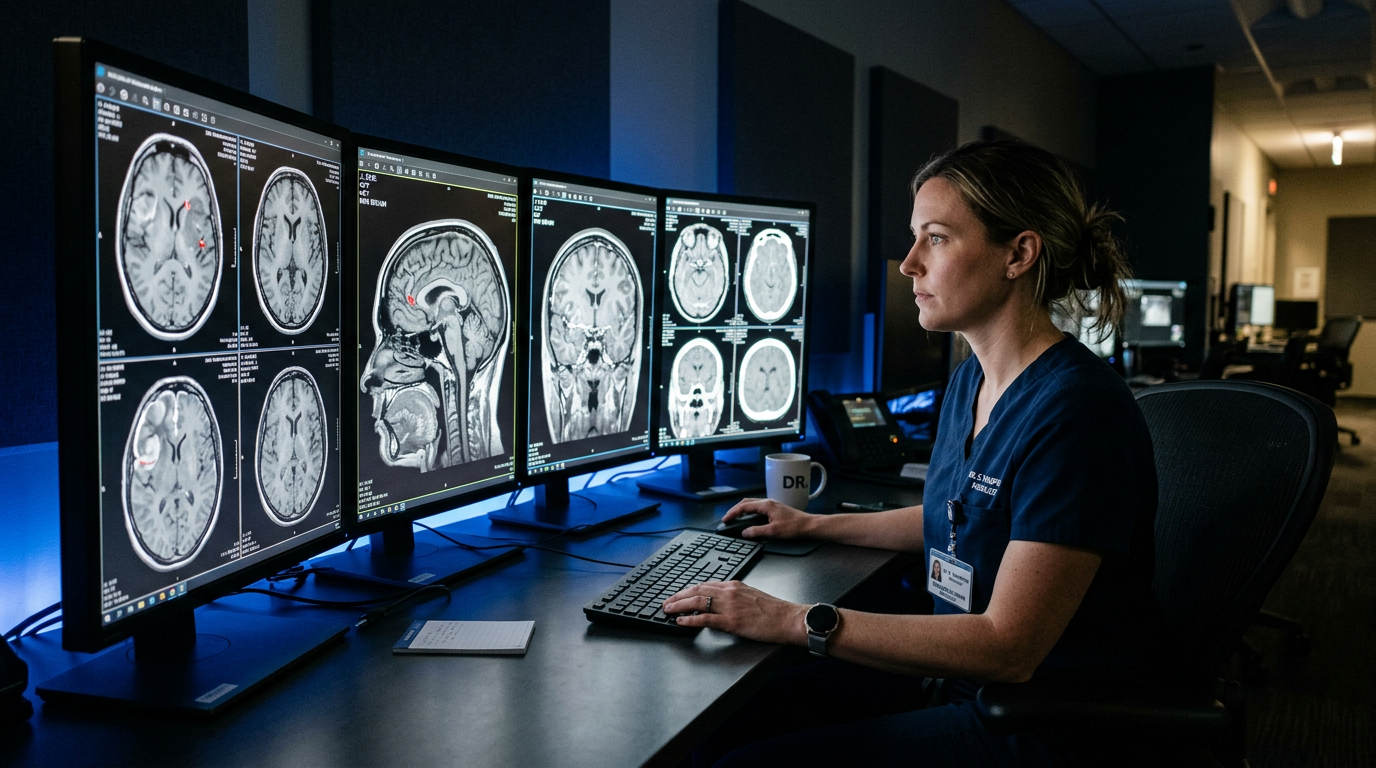
Artificial intelligence (AI) is increasingly being used to help radiologists review brain scans. It does not replace doctors, but it may help flag possible aneurysms that deserve a closer look.[ninds.nih][jacr]
What Is a Brain Aneurysm?
A brain aneurysm happens when a weak area in an artery wall bulges outward. Some aneurysms remain small and stable, while others may grow or press on nearby nerves and brain tissue.
Risk factors can include smoking, high blood pressure, family history, and certain genetic conditions. The risk of rupture depends on several factors, including aneurysm size, location, growth over time, and a person’s overall health.[ninds.nih][ninds.nih]
Symptoms That Need Urgent Care
A ruptured brain aneurysm is a medical emergency. The most common warning sign is a sudden, severe headache—often described as “the worst headache of my life.”[ninds.nih][mayoclinic]
Call emergency services immediately if someone has:
- A sudden, extremely severe headache
- Nausea or vomiting with the headache
- Stiff neck or sensitivity to light
- Double vision or sudden vision changes
- Drooping eyelid or a widened pupil
- Weakness, numbness, confusion, seizure, or loss of consciousness[ninds.nih][ninds.nih]
Do not wait to see whether symptoms improve. Do not drive yourself to the hospital if a ruptured aneurysm is possible.
How Doctors Look for Brain Aneurysms
Doctors may use imaging tests such as CT angiography (CTA), MR angiography (MRA), or digital subtraction angiography to examine blood vessels in the brain. A CTA uses contrast dye and CT imaging to create detailed pictures of the arteries.
Radiologists review many image slices and look for unusual bulges. This can be challenging because brain arteries are complex, aneurysms may be small, and image quality can vary.[pmc.ncbi.nlm.nih][nature]
How AI Can Help Radiologists
AI tools for brain imaging can quickly scan CT angiograms and highlight areas that may contain an aneurysm. The radiologist then reviews the flagged area and decides whether it is a real finding, an anatomical variation, or a false alarm.
In a real-world prospective study of more than 3,800 CTA exams, an FDA-cleared AI tool identified 55 true aneurysm cases that had not been reported during the initial radiologist review. This was a 39% relative increase in detection compared with radiologist-only review.[jacr][neimanhpi]
The study also found:
- AI was more sensitive than radiologists alone, meaning it flagged more true aneurysms.
- Radiologists had a higher positive predictive value, meaning their positive findings were more likely to be correct.
- Both AI and radiologists missed some true aneurysms.[neimanhpi]
This is why AI is best viewed as a support tool, not a final diagnosis.
Why Human Review Still Matters
AI can help draw attention to possible findings, but it can also produce false positives. In the same study, 46 of 101 AI-only findings were false positives.[neimanhpi]
A radiologist considers much more than the image, including:
- The patient’s symptoms and medical history
- Image quality and scan technique
- Normal anatomical variations
- Whether a finding needs follow-up imaging or treatment
- The risks and benefits of further testing
AI performance can also vary with scan quality, scanner settings, reconstruction methods, and patient populations. FDA clearance does not guarantee that a tool will perform equally well in every hospital or with every type of scan.[pmc.ncbi.nlm.nih][nature]
Can AI Prevent Aneurysm Rupture?
AI may help detect an aneurysm earlier, but it cannot predict rupture with certainty or prevent one by itself. Finding an aneurysm allows a care team to assess whether monitoring, lifestyle changes, or treatment may be appropriate.
Possible next steps after detection may include:
- Observation and follow-up imaging for some small, stable aneurysms
- Blood-pressure control and smoking cessation, which may reduce some vascular risks
- Specialist referral to a neurosurgeon or interventional neuroradiologist
- Treatment, such as surgical clipping or endovascular coiling, in selected higher-risk cases
Treatment decisions are individualized. A small aneurysm is not always treated, and a larger aneurysm is not always treated immediately.[ninds.nih][ninds.nih]
Benefits and Limitations of AI
| Potential benefit | Important limitation |
| May flag possible aneurysms for closer review | Can create false-positive alerts [neimanhpi] |
| May support radiologists during busy workflows | Does not replace clinical judgment [pmc.ncbi.nlm.nih] |
| May help detect some small or subtle findings | Performance can vary with image quality and setting [nature] |
| May support earlier specialist review | More detection does not automatically prove better long-term outcomes [link.springer] |
Key Takeaway: Doctor-Style Recommendation
AI can be a useful second set of eyes for radiologists reviewing brain scans for aneurysms, but it does not replace expert human review. If you have a sudden severe headache, vision changes, weakness, confusion, seizure, or loss of consciousness, seek emergency care immediately. If an aneurysm is found, discuss monitoring and treatment options with a neurovascular specialist.
Editorial Note
This article is for general educational purposes only and is not medical advice. It does not diagnose, treat, cure, or prevent any disease. AI findings should always be interpreted by qualified radiologists and treating clinicians. If you may be experiencing a medical emergency, call your local emergency number immediately.
References
- Stein, S., et al. (2024). Real-world performance of an artificial intelligence algorithm for detection of intracranial aneurysms. Journal of the American College of Radiology.[jacr][neimanhpi]
- National Institute of Neurological Disorders and Stroke. Cerebral aneurysms.[ninds.nih][ninds.nih]
- Mayo Clinic. Brain aneurysm: Symptoms and causes.[mayoclinic]
- Choy, G., et al. (2023). Clinical applications of artificial intelligence in radiology. PMC / NIH.[pmc.ncbi.nlm.nih]
- Research on AI variability in intracranial aneurysm detection. Scientific Reports / Nature.[nature]
- Systematic review of AI in cerebral aneurysm imaging and clinical decision support. Journal of Digital Imaging.[link.springer]