The Eye as a Window to the Heart: Can Retinal Scans Predict AFib?
Medical Disclaimer: The information provided in this article is for educational and informational purposes only and does not constitute medical advice. Always consult a qualified healthcare provider regarding any medical condition or treatment.
Introduction
he medical community evaluates retinal scans to predict afib as an essential cardiovascular breakthrough. Traditionally, poets describe the human eye as the window to the soul. However, modern preventative medicine reveals a literal window into your heart.
Scientists recently discovered a powerful physiological link between delicate retinal tissue and cardiac arrhythmias. Specifically, deep-learning models detect atrial fibrillation risk directly through standard ophthalmic examinations.
Clinical trials establish that subtle retinal thinning flags arrhythmia risks years before standard symptoms emerge. Consequently, this predictive biomarker revolutionizes screening protocols for high-risk patients. Atrial fibrillation often evades standard detection during routine doctor visits. Furthermore, this silent condition remains a primary driver of ischemic stroke and heart failure, according to the American Heart Association.
Ophthalmology clinics already possess the necessary fundus photography technology worldwide. Therefore, clinicians can integrate these ocular screenings into regular eye health checkups. Indeed, researchers documented these microvascular patterns in a landmark study published by the British Journal of Ophthalmology. Ultimately, routine retinal photography offers a non-invasive shield against devastating cardiovascular events.
The Silent Threat of Atrial Fibrillation
To grasp the importance of this new finding, one must first understand the challenge posed by atrial fibrillation. AFib is the most common type of cardiac arrhythmia, or irregular heartbeat. It occurs when the heart’s two upper chambers, the atria, beat chaotically and out of sync with the two lower chambers, the ventricles. This disorganized electrical activity can cause a rapid, fluttering heartbeat, but for many, it causes no noticeable symptoms at all.
This “silent” nature is what makes AFib so perilous. The condition affects an estimated 50 million people worldwide, and its prevalence is rising. When the heart’s chambers don’t beat correctly, blood can pool and form clots. If a clot breaks free, it can travel to the brain and cause a devastating ischemic stroke. In fact, individuals with AFib are five times more likely to suffer a stroke than those with a regular heart rhythm. The disorder is also strongly linked to an increased risk of heart failure, chronic kidney disease, and even dementia.
The diagnostic dilemma is profound. Because symptoms like fatigue, shortness of breath, or heart palpitations can be mild, intermittent, or attributed to other causes, a huge number of cases go undetected. Some research indicates that over 60% of patients are unaware they have AFib until they are diagnosed, often after a major health event like a stroke. This underscores a critical need for new, accessible screening methods that can identify at-risk individuals proactively.
A New Frontier: Connecting Ophthalmology and Cardiology
The retina, a light-sensitive layer of tissue at the back of the eye, is a unique part of the human body. It is the only place where blood vessels and nerve tissue of the central nervous system can be directly visualized non-invasively. This makes it an invaluable resource for observing the real-time effects of systemic diseases on the microvasculature. For years, ophthalmologists have used retinal exams to spot signs of diabetes, high blood pressure, and high cholesterol. Now, cardiology may be the next field to benefit from this ocular window.
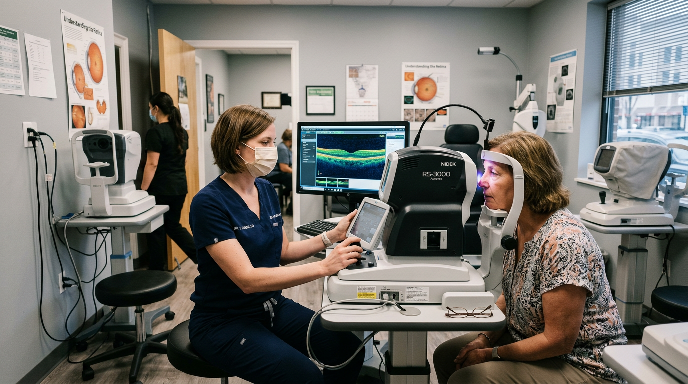
In a landmark investigation published in the journal *PLOS Digital Health*, a team of researchers explored whether structural changes in the retina could predict future AFib. The study leveraged two massive datasets: the UK Biobank and the AlzEye study, encompassing retinal images from over 90,000 individuals. The researchers used advanced imaging techniques, including Optical Coherence Tomography (OCT) and color retinal photography, to analyze the intricate layers of the retina.
OCT, in particular, was crucial to their work. It is a non-invasive imaging test that uses light waves to take cross-section pictures of the retina. Think of it as an “optical ultrasound,” allowing doctors to see each of the retina’s distinct layers and measure their thickness with micrometer precision.
Decoding the Retinal Clues
The investigation’s pivotal finding centered on a specific, measurable change within the retina. Individuals with a current or future diagnosis of AFib consistently exhibited a significant reduction in the thickness of two key retinal layers:
1. The Macular Ganglion Cell-Inner Plexiform Layer (mGCIPL):
This layer is composed of ganglion cells, which are specialized neurons that transmit visual information from the eye to the brain via the optic nerve.
2. The Inner Nuclear Layer:
This layer contains the cell bodies of various neurons that help process visual signals within the retina.
The thinning of the mGCIPL is believed to be a sign of retrograde neurodegeneration. The study’s authors hypothesize that this damage is caused by tiny, often silent, “mini-strokes” (microemboli) in the brain, a common and often undiagnosed consequence of AFib. These tiny clots disrupt blood flow, causing damage that works its way backward along the neural pathways to the ganglion cells in the retina, leading to their death and a subsequent thinning of the layer.
The thinning of the inner nuclear layer, however, suggests a different mechanism. The researchers propose this may be caused by direct damage from micro-strokes *within the eye itself*. The delicate vascular network of the retina is highly vulnerable to blockages, and the inner nuclear layer is particularly susceptible to this type of ischemic injury.
Most remarkably, the analysis revealed a powerful predictive element. Among participants who did not have AFib at the time of their eye scan, those who later went on to develop the condition already showed this characteristic retinal thinning. On average, these structural changes were detectable four years before the patient presented with AFib and received a formal diagnosis. This finding represents a monumental step forward, transforming the retinal scan from a diagnostic tool for existing disease into a predictive tool for future risk.
The Future of Integrated Health Screening
This research is more than just an interesting correlation; it points toward a future of more integrated and holistic healthcare. It reinforces the understanding that the body is an interconnected system, where the health of one part, like the circulatory system, is reflected in another, like the eye. This is a core principle of understanding overall Vascular Health & Circulation.
The implications for clinical practice are enormous. OCT scanners are already standard equipment in the offices of most ophthalmologists and many optometrists. If these findings are validated in further studies, incorporating an automated analysis of retinal layer thickness into routine eye exams could become a low-cost, non-invasive, and highly scalable screening method for AFib risk.
Imagine a future where your annual eye check-up does more than just update your glasses prescription. The data from your OCT scan could be analyzed by an AI algorithm trained to detect the subtle patterns of retinal thinning associated with cardiac risk. If a high-risk profile is detected, your optometrist or ophthalmologist could then refer you to your primary care physician or a cardiologist for further evaluation, such as an electrocardiogram (ECG) or extended heart rhythm monitoring.
This approach embodies the essence of a major Clinical Breakthrough: it takes existing, safe technology and applies it in a novel way to solve a major public health problem. It shifts the paradigm from reactive treatment—managing AFib after a diagnosis or, worse, after a stroke—to proactive identification and prevention.
What This Means for You Today
While the prospect of using eye scans for heart-risk screening is exciting, it is not yet standard clinical practice. This research is a critical first step, and further validation is needed before it can be widely implemented. However, this study provides powerful takeaways for anyone concerned with their long-term health:
* Value Your Eye Exams:
Do not skip your regular appointments with an eye care professional. These exams are about far more than vision; they are a crucial check-up for your overall health.
* Know Your AFib Risk Factors:
Discuss your personal and family medical history with your doctor. Key risk factors for AFib include high blood pressure, diabetes, obesity, sleep apnea, excessive alcohol consumption, and advancing age.
* Recognize the Symptoms:
While often silent, AFib can cause symptoms. Be aware of sustained heart palpitations, a “fluttering” or “thumping” in your chest, unexplained fatigue, dizziness, lightheadedness, and shortness of breath. If you experience these, seek medical evaluation.
* Embrace a Heart-Healthy Lifestyle:
The best way to prevent AFib and other cardiovascular diseases is through lifestyle choices. Maintain a healthy weight, eat a balanced diet, engage in regular physical activity, limit alcohol, and do not smoke.
The eye is an extraordinary organ. This research adds another layer to its complexity, revealing it as a sensitive barometer of our systemic health. By looking deep into the retina, we may soon be able to see the future of our hearts and take action before it’s too late.
Editorial Note: This article was independently researched and written based on current publicly available health information and does not represent the views of, and is not affiliated with, any external publication referenced.
References
1. Huemer, J., et al. (2024). Retinal thinning as a predictive biomarker for atrial fibrillation: a prospective cohort study from UK Biobank and AlzEye. *PLOS Digital Health*. https://doi.org/10.1371/journal.pdig.0001661
2. Li, H., et al. (2024). Global, regional, and national burden of atrial fibrillation/flutter, 1990–2021: results from the Global Burden of Disease Study 2021. *European Heart Journal – Quality of Care and Clinical Outcomes*. https://www.ncbi.nlm.nih.gov/pmc/articles/PMC12673497/
3. Wolf, P. A., Abbott, R. D., & Kannel, W. B. (1991). Atrial fibrillation as an independent risk factor for stroke: the Framingham Study. *Stroke*. https://www.ahajournals.org/doi/10.1161/01.str.22.8.983