AI in Radiology: A New Digital Safety Net for Brain Aneurysm Detection
Medical Disclaimer: The information provided in this article is for educational and informational purposes only and does not constitute medical advice. Always consult a qualified healthcare provider regarding any medical condition or treatment.
Introduction
Deep within the intricate network of blood vessels that supply our brain, a silent and potentially deadly threat can form. A brain aneurysm—a small, balloon-like bulge in an artery wall—is a vascular weak spot that often develops without any symptoms. For the estimated 1 in 50 people living with an unruptured brain aneurysm, it can be a ticking time bomb. While most will never rupture, those that do can cause a catastrophic bleed known as a subarachnoid hemorrhage, a medical emergency with a high rate of mortality and long-term disability.
For decades, the primary line of defense against this hidden danger has been the sharp eye of a radiologist. Tasked with meticulously examining hundreds of images from a single computed tomography (CT) scan, these medical experts hunt for subtle abnormalities in the brain’s complex vascular architecture. It is a high-stakes, high-pressure job where a momentary lapse or a particularly small, well-hidden aneurysm can have profound consequences.
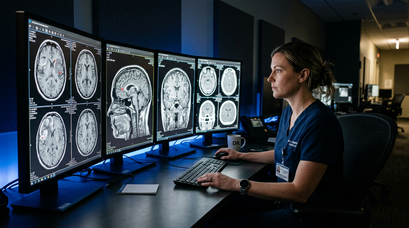
Now, a new technological partner is entering the reading room, offering a digital safety net to support these human experts. A landmark study has revealed the powerful potential of artificial intelligence (AI) to augment the skills of radiologists, creating a human-machine collaboration that significantly boosts the detection of these elusive aneurysms. This development marks a pivotal moment in the field of Vascular Health & Circulation, suggesting a future where technology helps catch what the human eye might miss.
The Silent Threat: Understanding Brain Aneurysms
To appreciate the significance of this technological advance, it’s essential to understand the nature of the threat. A brain aneurysm occurs when pressure from blood flow causes a weakened section of an artery wall to bulge outwards. Think of it as a weak spot on the inner tube of a bicycle tire that begins to protrude.
These aneurysms can be categorized into two main states:
1. Unruptured Aneurysms:
These are often small and asymptomatic. They are frequently discovered by chance during brain imaging for other conditions, such as chronic headaches, seizures, or following a head injury. While they may not pose an immediate danger, their discovery prompts a crucial decision-making process involving careful risk assessment.
2. Ruptured Aneurysms:
If the thinned wall of the aneurysm tears, it releases blood into the space surrounding the brain. This event, a subarachnoid hemorrhage, causes a sudden, excruciating headache often described as “the worst headache of my life,” and is a life-threatening emergency requiring immediate medical intervention.
Key risk factors for developing brain aneurysms include a family history of the condition, certain genetic disorders, untreated high blood pressure, and cigarette smoking. The challenge for clinicians has always been to identify at-risk individuals and, more importantly, to accurately detect existing aneurysms before they have a chance to rupture. This is where the field of diagnostic radiology plays a central role.
The Radiologist’s Burden: Finding a Tiny Flaw in a Complex System
When a brain aneurysm is suspected, the gold standard for non-invasive imaging is often a CT angiogram (CTA). This procedure involves injecting a contrast dye into the bloodstream, which illuminates the arteries on a CT scan, providing a detailed 3D map of the brain’s vascular tree.
The resulting dataset can contain hundreds, sometimes thousands, of individual image slices that a radiologist must painstakingly review. They scroll through the images, mentally reconstructing the twisting and turning pathways of the arteries, looking for any abnormal outpouching. It’s a task that requires immense concentration and expertise. However, the reality of modern medicine includes heavy workloads, the need for rapid interpretations in emergency settings, and the simple fact of human fallibility. Small aneurysms, particularly those under 3 millimeters, or those located in anatomically complex areas, can be incredibly difficult to spot.
This is the precise clinical gap that AI is poised to fill—not by replacing the radiologist, but by serving as an tireless, ever-vigilant digital assistant.
A Powerful Partnership: New Research Highlights a Collaborative Future
A groundbreaking prospective study published in the *Journal of the American College of Radiology* has provided compelling real-world evidence of this new collaborative model. Researchers at Northwell Health evaluated an FDA-cleared AI algorithm designed to detect intracranial aneurysms on CTA scans.
In the study, which included over 3,800 examinations, the AI software analyzed the scans in parallel with the standard clinical workflow. Critically, the radiologists performing the initial interpretations were “blinded” to the AI’s findings. This allowed the researchers to directly compare the performance of the human expert alone versus the potential of a human-AI team.
The results were striking. The AI algorithm successfully identified 55 genuine aneurysms that had been missed during the initial radiological review. This represented a 39% relative increase in detection—a significant improvement that could have life-altering implications for patients.
The study beautifully illustrated the complementary strengths of human and machine intelligence:
* AI’s High Sensitivity:
The AI algorithm demonstrated a higher sensitivity than radiologists alone (84.6% vs. 71.8%). In simple terms, this means the AI was better at “casting a wide net” and flagging any potential abnormality that could be an aneurysm. It was less likely to miss a true finding.
* Radiologist’s High Specificity:
Conversely, radiologists had a higher positive predictive value (92.7% vs. 78.2%). This means that when a radiologist definitively identified a finding as an aneurysm, they were more likely to be correct than the AI. The human expert excelled at filtering out “false positives” and applying clinical judgment to confirm a diagnosis.
This dynamic creates an ideal partnership. The AI acts as a first-pass screening tool, flagging suspicious areas that warrant a second look. The radiologist then applies their deep medical knowledge and experience to verify the AI’s findings, distinguish true aneurysms from anatomical variants, and place the discovery in the context of the individual patient’s health. It is a system of checks and balances that leverages the best of both worlds.
The Importance of Finding the Smallest Flaws
One of the most crucial findings from the study was the AI’s proficiency in detecting very small aneurysms—the very ones most likely to be overlooked by the human eye during a busy shift. While a rupture is often associated with larger aneurysms, the decision to monitor or treat an aneurysm is complex. It isn’t based on size alone, but also on its shape, its location, and the patient’s overall risk profile.
Identifying a small aneurysm early on provides a critical window of opportunity. It allows the clinical team and the patient to make informed decisions. This might involve:
* Active Surveillance:
For many small, stable aneurysms, the risk of intervention may outweigh the risk of rupture. In these cases, the best course of action is often watchful waiting, with periodic imaging to ensure the aneurysm is not growing or changing shape.
* Lifestyle Modification:
The discovery can serve as a powerful motivator for patients to address modifiable risk factors like quitting smoking or managing high blood pressure, which can slow aneurysm growth.
* Proactive Treatment:
If a small aneurysm has high-risk features (e.g., an irregular shape or a critical location) or if the patient has other risk factors (like a family history of rupture), a neurosurgeon may recommend proactive treatment. This can include surgical clipping or less-invasive endovascular coiling to secure the aneurysm and prevent a future bleed.
By flagging these small lesions, AI empowers a more proactive and personalized approach to Preventative Medicine, shifting the paradigm from reacting to a rupture to preventing one in the first place.
The Bigger Picture: Augmentation, Not Replacement
This study demonstrates a wider shift across modern clinical practice. Healthcare systems now integrate artificial intelligence to augment, rather than replace, physician expertise. Consequently, anxiety over machine replacement is diminishing rapidly. Clinicians now view algorithmic tools as powerful performance multipliers. Ultimately, these clinical breakthroughs are redefining standard diagnostic capabilities.
Today, engineers train specialized algorithms to manage massive datasets and complex visual patterns. Key examples include:
Identifying subtle, early-stage lung nodules on low-dose chest CTs.
Detecting acute ischemic or hemorrhagic stroke signals on emergency brain MRIs.
Measuring volumetric arterial plaque accumulation inside coronary blood vessels.
Offloading this repetitive perceptual work directly mitigates physician burnout. Furthermore, it restores cognitive bandwidth for higher-level patient care. Specialists gain more time to consult colleagues, guide nervous patients, and map intricate treatment protocols.
However, widespread clinical integration still faces notable friction. Hospital networks must resolve workflow bottlenecks, preserve patient data privacy, and validate software across diverse demographic populations.
Additionally, researchers must confirm whether higher detection rates truly improve long-term patient outcomes. Clinicians also remain vigilant regarding overdiagnosis risks. Flagging harmless, incidental lesions can trigger unnecessary diagnostic procedures and severe psychological distress.
Nevertheless, overwhelming clinical evidence points toward a collaborative future. This digital co-pilot works quietly in the background. It consistently boosts diagnostic precision, accelerates early detection, and protects patient safety before silent emergencies escalate.
Editorial Note: This article was independently researched and written based on current publicly available health information and does not represent the views of, and is not affiliated with, any external publication referenced.
References
1. Stein, S., et al. (2024). Real-World Performance of an Artificial Intelligence Algorithm for Detection of Intracranial Aneurysms. *Journal of the American College of Radiology*. https://doi.org/10.1016/j.jacr.2024.03.019
2. National Institute of Neurological Disorders and Stroke. (2023). *Cerebral Aneurysms Fact Sheet*. Retrieved from https://www.ninds.nih.gov/health-information/disorders/cerebral-aneurysms
3. Greving, J. P., et al. (2014). The PHASES score for prediction of risk of rupture of intracranial aneurysms: a pooled analysis of six prospective cohort studies. *The Lancet Neurology*, 13(1), 59-66. https://doi.org/10.1016/S1474-4422(13)70263-1